A Topographical Approach to Early Cancer and Connective Tissue Disease Detection
Earlier detection of cancer and connective tissue disorders may be possible using atomic force microscopy.
Tune in to learn how it works and discover:
The advantages of atomic force microscopy over electron microscopy
How fibroblasts use collagen to travel throughout the body
How fibrosis occurs and why it matters
Atomic force microscopy is a technique that can generate high-resolution images of biological samples too small to be imaged using other techniques.
It works similar to a stylus on a record player: a tiny stylus contacts and moves up and down along the biological sample, while a computer program analyzes these movements and recreates 3D images accordingly.
In essence, it’s a process whereby the topography of almost any surface can be measured and visualized.
This technology is leading to great insights in various fields of research, and has exciting applications in the field of early cancer detection.
Press play for all the details.
Episode also available on Apple Podcast: http://apple.co/30PvU9C
-
 0:45
0:45
FGP
3 days ago📚✊ Unraveling China's Cultural Revolution Mystery 🇨🇳 👀
56 -
 4:55
4:55
KTNV
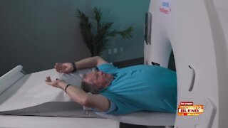
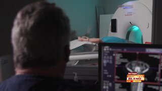
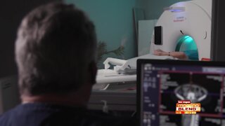
3 years agoEarly Detection in Pancreatic Cancer
48 -
 4:17
4:17
WXYZ
2 years agoEarly Signs of Alzheimer's Disease
95 -
 6:31
6:31
WMAR
3 years agoMedStar Health Cancer Network - Early Detection
61 -
 6:11
6:11
KTNV
2 years agoEarly Detection Could Save Your Life!
116 -
 4:19
4:19
KTNV
2 years agoThe Importance Of Mammograms & Early Detection
7 -
 5:46
5:46
KTNV
2 years agoEarly Detection Could Save Your Life
40 -
 5:40
5:40
KTNV
2 years agoEarly Detection Could Save Your Life
22 -
 5:05
5:05
KTNV
2 years agoEarly Detection To Save Your Life!
22 -
 3:06
3:06
WMAR
3 years agoColonoscopy helps detect cancer early
36